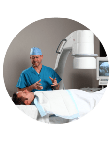
RP Eagle can provide your facility with comprehensive support for existing or future interventional radiology programs. Our Board Certified Interventional Radiologists will work with your clinical staff to enhance the quality of the patient experience and your facility’s interventional radiology program. Interventional radiology is a sub-specialty which uses a variety of imaging techniques to diagnose disease. Interventional radiology often uses minimally invasive procedures to eliminate the need for large incisions, which means less pain and quicker recovery for patients.
Angiography
Angiography is a medical technique used to visualize the inside of blood vessels or organs. A radiologist will inject a contrasting agent into the vessel before using x-ray to take images of the area. This technique can be used to view vessels in the heart, chest, lungs, kidneys, brain, neck, limbs or gastrointestinal tract.
Vascular Intervention
Many conditions which once required surgery can now be treated without surgery thanks to interventional radiologists. Vascular interventions include treatment of varicose veins, deep vein thrombosis, pulmonary embolism or acute limb ischemia.

Biliary Intervention
Many conditions which once required surgery can now be treated without surgery thanks to interventional radiologists. Vascular interventions include treatment of varicose veins, deep vein thrombosis, pulmonary embolism or acute limb ischemia.
GU/GI Intervention
Our interventional radiologists use a variety of imaging tools to see internal structures of the GU and GI tract. Treatment is delivered through catheters or other tiny instruments which are inserted through very small incisions or natural pathways. Interventional radiologists specialize in guiding these instruments through the body using the latest imaging techniques.
Myelography
Myelography is a type of imaging exam which uses a contrast medium to detect problems in the spinal cord. Radiologists use this type of imaging to locate a spinal cord injury, cyst or tumor. One of the most common uses of myelogram is to help find the cause of back pain when MRI or CT has been unsuccessful in locating the problem. Myelography is helpful with surgical planning when patients are prohibited from having an MRI because of screws, plates of rods in the spine.

Biopsy
Samples of tissue may be needed to indentify the cause of certain diseases. Radiologists use imaging guidance to remove a tissue sample from an area of interest for pathological examination. Interventional radiologists may use a small needle to remove tissue samples.
THE SERVICES LISTED ON THIS WEBSITE ARE FOR GENERAL INFORMATION PURPOSES ONLY AND DO NOT INCLUDE ALL SERVICES OF RP EAGLE. WHILE WE STRIVE TO KEEP THE INFORMATION UP TO DATE AND CORRECT, WE MAKE NO REPRESENTATIONS OR WARRANTIES OF ANY KIND, EXPRESS OR IMPLIED, ABOUT THE CONTENT, COMPLETENESS, ACCURACY, RELIABILITY, LEGALITY, SUITABILITY OR AVAILABILITY, WITH RESPECT TO THE SERVICES CONTAINED ON THIS WEBSITE.
